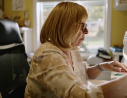

Dr. Cherie Cora hopes women see dense breast information not as a warning, but as an invitation to be proactive
Breast density may not be something most women think about until they read it on their mammogram report. For many women, it’s an important factor that can influence both cancer risk and the sensitivity of screenings.
Cherie Cora, MD, director of breast imaging and radiologist at Loma Linda University Cancer Center, explains that breast density refers to the amount of fibroglandular tissue compared to fat in the breast.
But why does this matter?
On a mammogram, fat appears dark while normal breast tissue and potential cancers appear white. “In a fatty breast, it’s like looking for stars in a dark sky, easy to spot. But in a dense breast, everything blends together,” Cora says.
Normal dense tissue and abnormalities appear on a scan in the same shade; therefore, small cancers can sometimes be difficult to find. That’s why many women with dense breasts ask about additional imaging like ultrasound or MRI. Yet, Cora says that decision should be individualized.
“Ultrasound can help us look through dense tissue, but it also increases the chance of finding something that requires follow up or biopsy that ultimately turns out to be benign,” she says. “It’s a balance between vigilance and avoiding unnecessary anxiety.”
Since federal and state law requires radiology centers to notify patients if they have dense breast tissue, many women now see that information in their results letter. Cora explains that if your report says your breasts are classified as heterogeneously dense or extremely dense, that doesn’t mean something is wrong. It just means you should discuss it with your provider and see if additional screening makes sense for your individual risk.
For most average-risk women, mammography remains the cornerstone of early detection. “Dense breasts don’t make mammograms useless,” Cora emphasizes. “We can still see calcifications and other signs of early disease.”
Advancements in imaging help radiologists better visualize dense tissue. One of the biggest breakthroughs, Cora says, is 3D mammography, also known as tomosynthesis.
“Instead of taking one flat picture, the machine moves in an arc and captures multiple slices through the breast,” she explains. “That separation helps us distinguish overlapping structures and identify subtle distortions or small masses that might otherwise be hidden.”
Other technologies, such as contrast-enhanced mammography or nuclear medicine imaging are also emerging, but Cora says mammography remains the gold standard for screening.
For women wondering when to start, Cora follows the guidance of the American College of Radiology: annual mammograms beginning at age 40 for women at average risk.
“All women, in particular Black and Ashkenazi Jewish women, should have a formal breast cancer risk assessment by age 25,” says Cora. “If their lifetime risk is higher than 20 percent, they might begin mammographic screening earlier and/or add MRI.”
Cora also emphasizes the importance of continuing regular screenings later in life. “People are living longer, and cancer can still develop in your 70s, 80s, and beyond,” she says. “If you’re healthy enough to treat it, you’re healthy enough to keep screening.”
Cora hopes women see dense breast information not as a warning, but as an invitation to be proactive. “Knowing your breast density helps you make informed decisions,” she says. “Talk to your doctor. Get your screening mammograms and don’t abandon them, they’re still the primary and most accessible tool we have for catching cancer early, when it’s most treatable.”
Schedule your mammogram or a breast health consultation at Loma Linda University Cancer Center.
More stories about: Breast Cancer Cancer Center Primary Care Healthcare Women's Health
Related Articles