The Loma Linda University Department of Urology has added an MRI and ultrasound fusion system for prostate biopsies, the latest development on the cutting edge of prostate cancer care.
The new method of fusing MRI and ultrasound images improves the location accuracy of prostate biopsies, which can minimize the number of biopsies needed for the patient and give doctors better insight on how to treat the cancer. The new approach helps doctors identify dangerous cancers to treat aggressively and allows them to monitor low-risk cancer with an active surveillance strategy.
Urology specialist Herbert C. Ruckle, MD,chair of Loma Linda University Health Urology Department, says having the technology can reassure patients in the region that they have access to cutting edge and comprehensive prostate cancer care.
“This keeps Loma Linda University Health at the forefront of where the specialty is at in terms of the detection and management of localized prostate cancer,” Ruckle said. “Patients can have confidence in knowing they are receiving state-of-the-art care.”
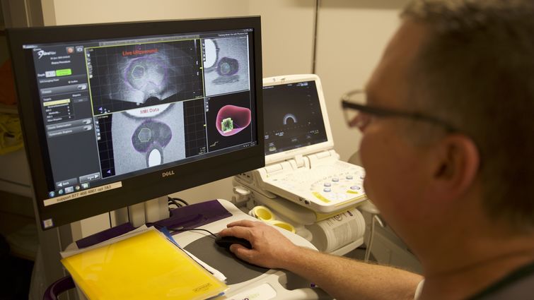
MRI is currently the most accurate method of imaging the prostate. An MRI/ultrasound fusion biopsy now uses images from images of the prostate from the previously obtained MRI and the real time ultrasound images to guide the prostate biopsy at areas suspicious for cancer. First, a radiologist reads an MRI of the prostate and marks the boundaries of the prostate and any suspicious lesions on the MRI pictures. Those images are then sent to a computer in the Urology Department. A urologist then starts the biopsy procedure using an ultrasound, then overlays the marked MRI image on the ultrasound image to pinpoint precisely where the lesions are. A biopsy needle is then directed to the suspicious area with greater precision than previously possible.
The current prostate biopsy method typically used in urology practice only utilizes transrectal ultrasound imaging during a biopsy. An ultrasound approach by itself has a less detailed image and doesn’t offer the accuracy or high resolution of an MRI regarding the location of the lesion to be biopsied. This typically results in patients needing more biopsies to find the cancer. It can also delay patients with more aggressive forms of cancer from being treated immediately because of the difficulty in finding the cancer.
Ruckle says that having a clearer and more detailed picture of a patient’s prostate as it relates to the size and location of the cancer area improves cancer staging accuracy and equates to a more customized prostate cancer care with better outcomes.
“We are constantly trying to identify which men have dangerous prostate cancers needing aggressive treatment versus which patients have prostate cancers best managed by ongoing monitoring, and this fusion technique helps us do that,” Ruckle said. “Ultimately, our goal at Loma Linda University Health is to improve cancer outcomes for every person we treat.”
Physicians and staff place their patient’s overall health as a top priority at Loma Linda University Cancer Center. In addition to biopsies and cancer care, the center can point patients in the direction of resources to enhance whole person care. To learn more about the Cancer Center, visit the prostate cancer care website or call 1-800-782-2623.