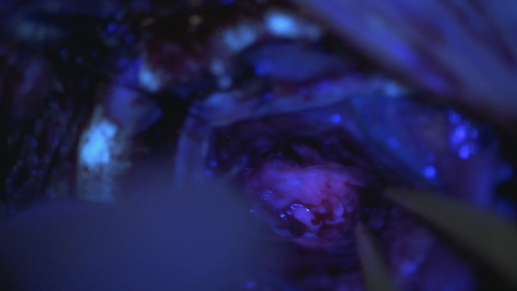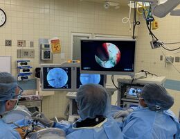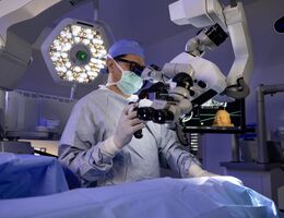
This photo shows the fluorescent features in gliomas under blue light of fluorescent-guided neurosurgery in a procedure performed by Dr. Tarek El Ahmadieh.
Brain surgery and the circumstances surrounding can leave patients with nerves and not much curiosity on operation day. But understanding the technological advances may relieve some stress. Whether you or a loved one have had or will be receiving treatment for high-grade gliomas, fluorescent-guided surgery ensures that your surgeon cleans more tumor cells.
Studies show that gliomas make up more than a quarter of all primary brain tumors. Surgical resection of them remains the first line of treatment. Fluorescence-guided surgery allows neurosurgeons, like Tarek El Ahmadieh, MD, at Loma Linda University Health, to have improved visualization of tumor cells in the operating room.
How does it work?
On the morning of surgery, patients drink a solution of 5-aminolevulinic acid (5-ALA). 5-ALA is a naturally occurring amino acid in the body and accumulates in glioma cells. The progression of 5-ALA presence in the tumor cells converts into having fluorescent features, creating a distinct visual difference between tumor and normal brain tissue for El Ahmadieh. The glow is only seen with a blue light. The blue light highlights tumor tissue as bright pink and leaves surrounding normal tissue as shadowy and violet.
El Ahmadieh says he cleans out as much of the glioma as possible, then turns on the blue light to show any leftover tumor tissue. This practice allows surgeons to clean out tumorous cells that aren’t visible to the naked eye. El Ahmadieh says some gliomas have infiltrative natures and spread far beyond areas detected on MRIs.
“To us surgeons’, surgical resection is an everyday practice that has shown to correlate positively with progression-free survival and quality of life,” El Ahmadieh said. “I think showing patients and their families how effective and advanced our technology is can provide some peace of mind.”
LLUH neurosurgeons provide the latest innovative surgical options for disorders of the brain, spine, and peripheral nerves. Learn more about brain tumor services and the conditions we treat.


