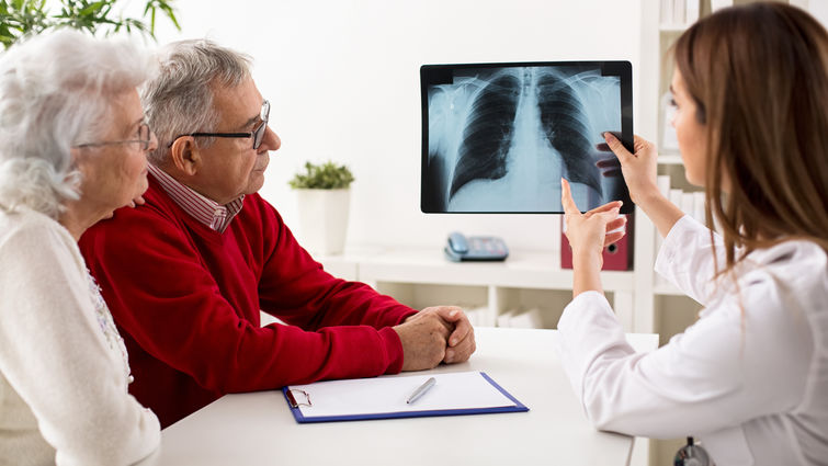
Interventional pulmonologists at Loma Linda University Health have performed thousands of minimally invasive procedures to diagnose and stage lung cancer. This Lung Cancer Awareness Month, one such expert imparts insights on advanced diagnostic bronchoscopy (ADB) and how its minimally invasive techniques smoothen the diagnostic experience for patients.
“Loma Linda University Health has consistently been the first institution in the Inland Empire to acquire technology and implement the practice infrastructure to offer our patients state-of-the-art, comprehensive lung cancer care,” says Ara Chrissian, MD, FCCP, DAABIP, director of Adult Bronchoscopy and Interventional Pulmonology at Loma Linda University Health
Of those technologies that have improved lung cancer diagnostic efforts, ADB enables physicians to biopsy the main mass or tumor in the lung and also determine the cancer’s stage by examining lymph glands in the center of the chest at the same time. Achieving both diagnosis and stage in a single procedure maximizes accuracy, minimizes risk, and allows patients to start treatment as soon as possible, Chrissian says.
ADB is a relatively straightforward and seamless process for patients, he says, with physicians performing the procedure in about 45-60 minutes while the patient is under sedation. During that time, the physician inserts a thin, long, and flexible tube with a tiny camera, called a bronchoscope, into the patient’s mouth, guiding the tool toward the primary tumor in the patient’s lung.
Physicians navigate precisely toward the tumor via a selection of three guidance techniques. These are often used together to provide maximum value:
- Endobronchial ultrasound: The bronchoscope has a video camera with an ultrasound probe attached to it that creates real time images of the lungs and lymph nodes. This allows for precise and accurate biopsy.
- Electromagnetic navigation: Functioning like a ‘GPS’ for the lungs, this specialized software digitally identifies targets in the lungs using images from CT scans. Physicians then guide a bronchoscope to the target.
- Robotic technology: This technology represents the next generation in precision. The robot drives the bronchoscope to the target using special technology. This provides the physician safe and accurate access to nodules that are both very small and on the very edge of the lung. Loma Linda University Health is currently one of the few specialized centers in the country to use this technology.
Physicians wield the bronchoscope the same way for all three techniques. Once the bronchoscope has located and reached the area of interest, the physician inserts biopsy tools — like a needle and small forceps — through the tube, then deploys them to collect several small pieces of lung tissue. Next, the tissue travels to a lab for analysis under a microscope by a pathologist.
While taking a biopsy of the lung’s primary tumor yields a lung cancer diagnosis, also taking a biopsy of nearby lymph glands in the chest provides important insights into the cancer’s staging, Chrissian says. For example, if the biopsy of the lymph glands reveals cancerous cells, physicians know the lung cancer has metastasized or spread and is thus at a later stage — this may require a different approach to therapy.
Again, as far as the patient is concerned, Chrissian says this process is instantaneous and painless. A patient remembers only the preparatory measures — not eating the day of the procedure, possibly pausing blood thinners, coming in a couple of hours early for IVs — and the short recovery period. This entire outpatient procedure takes place over a half-day, Chrissian says, and patients may resume their normal activities within several hours of leaving the hospital and attend work the next day.
Before minimally invasive techniques, physicians performed lung cancer diagnosis and staging via surgical procedures, such as mediastinoscopies, thoracoscopies, and even thoracotomies. Chrissian says each of these procedures is more invasive, involving incisions in the skin and chest and usually requiring hospitalization. ADB does not require any incisions.
Beyond aesthetic concerns, ADB also affords patients numerous other benefits, says Chrissian, including:
- Accuracy: During ABD, the physician is likely to acquire both the diagnosis and the stage all in one procedure. Surgical means of diagnosing lung cancer, though also accurate, usually require separate sessions of invasive procedures to obtain both a diagnosis and the cancer's staging.
- Safety: Less than 1% of individuals who undergo ADB require a hospital stay, whereas a hospital stay is a norm after a surgery.
- Reduced discomfort: At most, patients could experience a slight sore throat or a minor cough that ceases after a day.
Because ADB technology enables interventional pulmonologists like Chrissian to achieve diagnosis and staging in a single sitting, surgeons can focus on performing larger-scale procedures and surgeries. Moreover, the diagnostic test’s efficiency allows oncologists to receive more information about the patient’s condition upfront and respond as quickly as possible to provide the patient with the care they need.
“Advanced diagnostic bronchoscopies are easy, safe, efficient, and high-yield,” Chrissian says. “We look forward to continuing to provide patients with the care they need earlier and accurately."
To learn more about options for lung cancer screening or book an appointment, please visit the interventional pulmonology page or call 909-558-2896.